Endoscopic mucosal resection
Endoscopic mucosal resection is a procedure that uses an endoscope to remove carcinoma in situ and early-stage cancer from the lining of the digestive tract. An endoscope is a thin, tube-like instrument with a light and a lens and tools to remove tissue.
Surgery
Surgery is a common treatment for stomach cancer. The type of surgery depends on where the cancer is located.
Other treatments may be given in addition to surgery:
- Treatment given before surgery is called preoperative therapy or neoadjuvant therapy. Chemotherapy may be given before surgery to shrink the tumor and reduce the amount of tissue that needs to be removed during surgery. Chemoradiation given before surgery, to shrink the tumor, is being studied.
- Treatment given after surgery, to lower the risk that the cancer will come back, is called adjuvant therapy. After the doctor removes all the cancer that can be seen, some patients may be given chemotherapy, radiation therapy, or both to kill any cancer cells that are left.
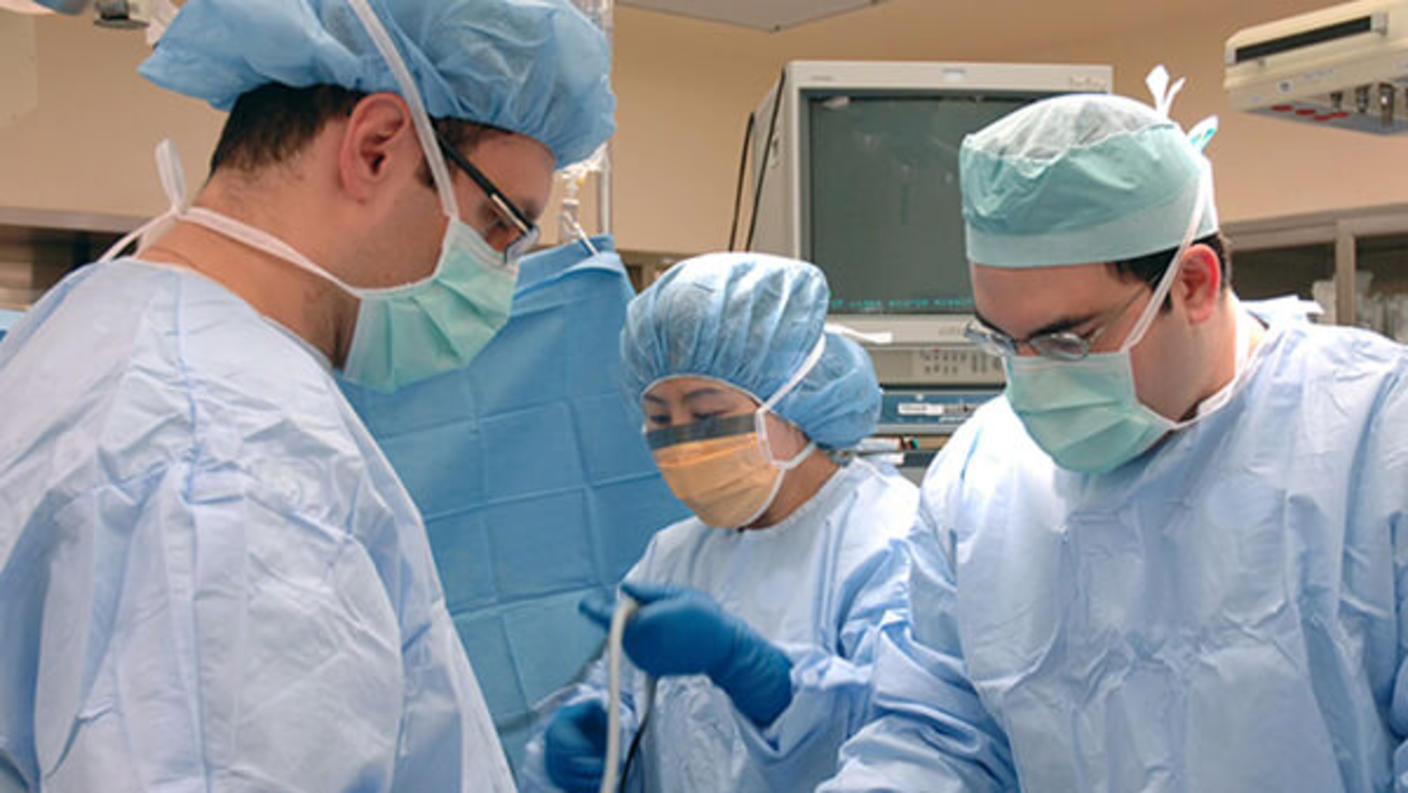
Gastrectomy
Gastrectomy, the removal of part or all of the stomach, is the main surgery for stomach cancer:
- Subtotal gastrectomy is the removal of the part of the stomach that contains cancer, nearby lymph nodes, and parts of other tissues and organs near the tumor. The spleen may also be removed.
- Total gastrectomy is the removal of the entire stomach, nearby lymph nodes, and parts of the esophagus, small intestine, and other tissues near the tumor. The spleen may also be removed. Then the surgeon attaches the esophagus to the small intestine so the patient can continue to eat and swallow.
Endoluminal stent placement
Endoluminal stent placement may be done when the tumor blocks the passage into or out of the stomach. In this procedure, the surgeon places a stent (a thin, expandable tube) from the esophagus to the stomach or from the stomach to the small intestine to allow the patient to eat normally.
Endoluminal laser therapy
Endoluminal laser therapy is a procedure in which an endoscope (a thin, lighted tube) with a laser attached is used as a knife to open a gastrointestinal blockage.
Gastrojejunostomy
Gastrojejunostomy is the removal of the part of the stomach with cancer that is blocking the opening into the small intestine. Then the surgeon connects the stomach to the jejunum (a part of the small intestine) to allow food and medicine to pass from the stomach into the small intestine.
Radiation therapy
Radiation therapy uses high-energy x-rays or other types of radiation to kill cancer cells or keep them from growing. Stomach cancer is sometimes treated with external radiation therapy. This type of radiation therapy uses a machine outside the body to send radiation toward the area of the body with cancer.
Learn more about External Beam Radiation Therapy for Cancer and Radiation Therapy Side Effects.
Chemotherapy
Chemotherapy (also called chemo) uses drugs to stop the growth of cancer cells, either by killing the cells or by stopping them from dividing.
Chemotherapy for stomach cancer is usually systemic, meaning it is injected into a vein or given by mouth. When given this way, the drugs enter the bloodstream to reach cancer cells throughout the body.
Chemotherapy drugs used to treat stomach cancer include:
- capecitabine
- cisplatin
- docetaxel
- doxorubicin
- epirubicin
- fluorouracil (5-FU)
- irinotecan
- leucovorin
- oxaliplatin
- paclitaxel
- trifluridine and tipiracil
To learn more about how chemotherapy works, how it is given, common side effects, and more, visit Chemotherapy to Treat Cancer and Chemotherapy and You: Support for People With Cancer.
Targeted therapy
Targeted therapy uses drugs or other substances to identify and attack specific cancer cells. Your doctor may suggest biomarker tests to help predict your response to certain targeted therapy drugs. Learn more about Biomarker Testing for Cancer Treatment.
Targeted therapies used to treat stomach cancer include:
Learn more about Targeted Therapy to Treat Cancer.
Immunotherapy
Immunotherapy helps a person’s immune system fight cancer. Your doctor may suggest biomarker tests to help predict your response to certain immunotherapy drugs. Learn more about Biomarker Testing for Cancer Treatment.
Immunotherapy drugs used to treat stomach cancer include:
These drugs work in more than one way to kill cancer cells. They are also considered targeted therapy because they target specific changes or substances in cancer cells.
Learn more about Immunotherapy to Treat Cancer and Immunotherapy Side Effects.
Hyperthermic intraperitoneal chemotherapy (HIPEC)
Regional chemotherapy is a method of placing chemotherapy directly into an organ or a body cavity, such as the abdomen, to mainly affect cancer cells in those areas.
A type of regional chemotherapy called hyperthermic intraperitoneal chemotherapy, or hot chemotherapy, is being studied to treat stomach cancer and may be offered at certain treatment centers. After the surgeon has removed as much of the cancer as possible during surgery, a chemotherapy drug, such as mitomycin or cisplatin, is warmed and pumped directly into the peritoneal cavity through a thin tube for about 2 hours. The surgeon then drains the chemotherapy from the abdomen and rinses the abdomen before closing the incision.
Clinical trials
A treatment clinical trial is a research study meant to help improve current treatments or obtain information on new treatments for patients with cancer. For some patients, taking part in a clinical trial may be an option.
Use our clinical trial search to find NCI-supported cancer clinical trials that are accepting patients. You can search for trials based on the type of cancer, the age of the patient, and where the trials are being done. Clinical trials supported by other organizations can be found at ClinicalTrials.gov.
To learn more, visit Clinical Trials Information for Patients and Caregivers.
Follow-up testing
Some tests that were done to diagnose or stage the cancer may be repeated to see how well the treatment is working. Decisions about whether to continue, change, or stop treatment may be based on the results of these tests. These tests are sometimes called follow-up tests or check-ups.
You may also have blood tests for tumor markers such as CEA and CA 19-9. Increased levels of these markers may mean your stomach cancer has come back. Learn more about Tumor Markers.