Studying Human Cancer Invasion and Metastasis in Real-Time in the Laboratory
, by Andrew J. Ewald, Ph.D. and Joel S. Bader, Ph.D.
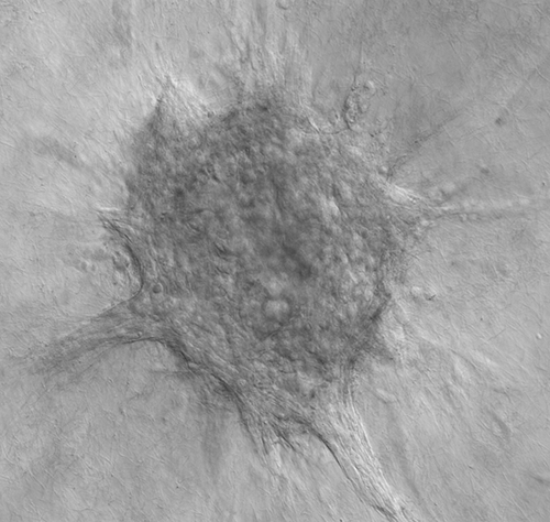
Human breast tumor organoid invades collectively into a 3D collagen I microenvironment.
A large majority of cancer deaths are attributable to metastasis, the process by which cancer cells spread throughout the body to form new tumors in distant vital organs1. Despite its central importance to patient outcomes, the cellular and molecular basis of metastasis is incompletely understood. Surprisingly, metastatic cancer has also been relatively understudied compared to earlier, more treatable stages of the disease2. This discrepancy may in part be attributable to metastasis involving the entire body, occurring deep inside the patient over long periods, and involving many different cells types and molecular pathways in complex combinations. These challenges come together to make metastasis difficult to study in the laboratory.
There has therefore been an urgent need to develop model systems that enable the analysis of key processes in metastasis at cellular and molecular resolution3. We set out to solve this problem inspired by progress with tissue organoid assays originally developed to study normal mammary gland development4,5. The essential concept is to surgically remove a portion of an organ, mechanically disrupt it with a scalpel, digest with enzymes, and then process by centrifugation to separate single cells from the tissue pieces or organoids. The single cells consist mostly of immune cells or fibroblasts and can be included or excluded from the culture as desired. Organoids usually consist of 100-500 epithelial cells. Our approach contrasts with the stem cell organoids pioneered by Hans Clevers: our tissue organoids are freshly isolated, are not generally expanded, do not require Wnt ligands for short term culture, and are available for immediate use. The basic approach of mechanical and enzymatic processing to generate tissue organoids is very flexible, and we have adapted it for use with normal mammary glands, primary breast tumors, and metastatic site breast tumors. We have also extended these methods to liver, pancreas, and lung tumors and can start from fresh human tumors, patient derived xenografts (PDXs), or genetically engineered mouse models (GEMMs). We routinely generate ~2,000 organoids per clinical sample, ~20,000 per PDX tumor, and ~200,000 per GEMM model. Accordingly, we can explant organoids from the same sample into a wide range of experimental conditions. We have applied this approach both to understand molecular pathways in the cancer cell and also to interrogate the functional role of specific elements of the tumor microenvironment6,7,8,9.

Andrew J. Ewald, Ph.D. and Joel S. Bader, Ph.D., professors at Johns Hopkins University
One of the main advantages of our approach is the ability to maintain a tumor's cellular and phenotypic diversity in convenient, multi-well plate compatible assays. Metastasis requires many steps, each of which poses quite distinct challenges for the cancer cell. Accordingly, we have developed a range of assays modeling primary tumor growth, invasion, dissemination, intravasation, and secondary tumor formation. We use highly automated microscopy to image the behavior of cancer cells at subcellular resolution in these assays, which enables us to develop hypotheses for how these processes take place at the cellular level. To understand the underlying molecular regulation, we can introduce signaling perturbations, shRNA, CRISPR, inducible gene expression, or Cre-lox based gene deletion. We have also optimized nucleotide and protein-based molecular analyses in these cultures, enabling mechanistic dissection of the molecular basis of phenotypes. We then integrate the results of the experimental interventions using network analysis and computational modeling. We have projects analyzing the molecular basis of normal, primary tumor, and metastatic site growth and motility that enable us to identify the truly cancer-specific processes.
Our newly launched Center for Cancer Target Discovery and Development (CTD²) builds on our track record of success and provides substantial new resources to exploit these model systems to develop novel therapeutic targets. Breast cancer is particularly challenging in exhibiting a very wide range of suspected driver mutations, with relatively few present in large percentages of patients10,11. Accordingly, the understanding of which genes are the most important for breast tumor initiation and particularly for metastasis remains largely incomplete. The key insight guiding our CTD² Center is that quantitative analysis of phenotypes within these 3D organoid assays enables us to adopt the mathematical framework of population genetics to identify genes and mutations responsible for breast cancer growth, invasion, and metastasis. The large number of organoids isolated, combined with quantitative imaging and applied mathematics, enables us to systematically analyze the molecular basis of variation among different clones within the same tumor and between tumors from different individuals. Based on our previous analyses of molecular programs driving collective invasion in breast cancer6 and our past work in population genetics12,13, we anticipate that our approach will enable us to appreciate the underlying molecular logic of metastasis. We will then use techniques from network analysis and graph theory14,15 to prioritize targets for intervention.
Our goals are to understand metastasis at the molecular level, to apply these insights to identify patients at the greatest risk of metastatic recurrence, and ultimately to improve patient outcomes by identifying new anti-metastatic therapies. Past and current support from the BCRF, the JKTG Foundation, and the National Cancer Institute has been critical to the development of these techniques and to their current application in translational cancer research.
References
- Siegel RL, Miller KD, Jemal A. Cancer statistics, 2016. CA: A Cancer Journal for Clinicians. 2016 Jan 1;66(1):7-30. (PMID: 26742998)
- Schoger J. VOICES:‘Changing the Landscape for People Living with Metastatic Breast Cancer’—New Report from the Metastatic Breast Cancer Alliance. Oncology Times. 2014 Nov 10;36(21):36-37.
- Shamir ER, Ewald AJ. Three-dimensional organotypic culture: experimental models of mammalian biology and disease. Nature Reviews Molecular Cell Biology. 2014 Oct;15(10):647-664. (PMID: 25237826)
- Ewald AJ, Brenot A, Duong M, Chan BS, Werb Z. Collective epithelial migration and cell rearrangements drive mammary branching morphogenesis. Developmental Cell. 2008 Apr 15;14(4):570-581. (PMID: 18410732)
- Nguyen-Ngoc KV, Shamir ER, Huebner RJ, Beck JN, Cheung KJ, Ewald AJ. 3D culture assays of murine mammary branching morphogenesis and epithelial invasion. In Tissue Morphogenesis. 2015 (pp. 135-162). Humana Press, New York, NY. (PMID: 25245692)
- Cheung KJ, Gabrielson E, Werb Z, Ewald AJ. Collective invasion in breast cancer requires a conserved basal epithelial program. Cell. 2013 Dec 19;155(7):1639-1651. (PMID: 24332913)
- Cheung KJ, Padmanaban V, Silvestri V, Schipper K, Cohen JD, Fairchild AN, Gorin MA, Verdone JE, Pienta KJ, Bader JS, Ewald AJ. Polyclonal breast cancer metastases arise from collective dissemination of keratin 14-expressing tumor cell clusters. Proceedings of the National Academy of Sciences. 2016 Feb 16;113(7):E854-863. (PMID: 26831077)
- Nguyen-Ngoc KV, Cheung KJ, Brenot A, Shamir ER, Gray RS, Hines WC, Yaswen P, Werb Z, Ewald AJ. ECM microenvironment regulates collective migration and local dissemination in normal and malignant mammary epithelium. Proceedings of the National Academy of Sciences. 2012 Sep 25;109(39):E2595-2604. (PMID: 22923691)
- Shamir ER, Pappalardo E, Jorgens DM, Coutinho K, Tsai WT, Aziz K, Auer M, Tran PT, Bader JS, Ewald AJ. Twist1-induced dissemination preserves epithelial identity and requires E-cadherin. Journal of Cell Biology. 2014 Mar 3;204(5):839-856. (PMID: 24590176)
- Lin J, Gan CM, Zhang X, Jones S, Sjöblom T, Wood LD, Parsons DW, Papadopoulos N, Kinzler KW, Vogelstein B, Parmigiani G. A multidimensional analysis of genes mutated in breast and colorectal cancers. Genome Research. 2007 Sep;17(9):1304-1318. (PMID: 17693572)
- Wood LD, Parsons DW, Jones S, Lin J, Sjöblom T, Leary RJ, Shen D, Boca SM, Barber T, Ptak J, Silliman N. The genomic landscapes of human breast and colorectal cancers. Science. 2007 Nov 16;318(5853):1108-1113. (PMID: 17932254)
- Bader JS, Sham P. Family-based association tests for quantitative traits using pooled DNA. European Journal of Human Genetics. 2002 Dec 3;10(12):870-878. (PMID: 12461696)
- Sham P, Bader JS, Craig I, O'Donovan M, Owen M. DNA pooling: A tool for large-scale association studies. Nature Reviews Genetics. 2002 Nov 1;3(11):862-871. (PMID: 12415316)
- Park Y, Bader JS. Resolving the structure of interactomes with hierarchical agglomerative clustering. BMC Bioinformatics. 2011 Dec;12(Suppl 1):S44. (PMID: 21342576)
- Qi Y, Suhail Y, Lin YY, Boeke JD, Bader JS. Finding friends and enemies in an enemies-only network: A graph diffusion kernel for predicting novel genetic interactions and co-complex membership from yeast genetic interactions. Genome Research. 2008 Dec 1;18(12):1991-2004. (PMID: 18832443)