
What Is Gallbladder Cancer?
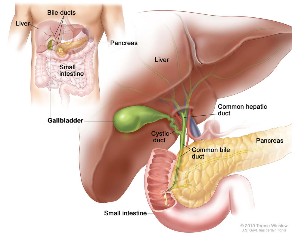
Anatomy of the gallbladder. The gallbladder is just below the liver. Bile is stored in the gallbladder and flows through the cystic duct and the common bile duct into the small intestine when food is being digested.
Credit: © Terese Winslow
Gallbladder cancer is a rare type of cancer that starts in the tissues of the gallbladder. Gallbladder cancer occurs when cells in the gallbladder start to grow out of control.
The gallbladder is a pear-shaped organ that lies just under the liver in the upper abdomen. The gallbladder stores bile, a fluid made by the liver to digest fat. When food is being broken down in the stomach and intestines, bile is released from the gallbladder through a tube called the common bile duct, which connects the gallbladder and liver to the first part of the small intestine.
The wall of the gallbladder has 4 main layers of tissue:
- mucosal (inner) layer
- muscle layer
- connective tissue layer
- serosal (outer) layer
Primary gallbladder cancer starts in the inner layer and spreads through the outer layers as it grows.