Tests to stage anal cancer
After anal cancer has been diagnosed, tests are done to find out if cancer cells have spread within the anus or to other parts of the body.
The process used to find out if cancer has spread within the anus or to other parts of the body is called staging. The information gathered from this staging process determines the stage of the disease. It is important to know the stage in order to plan treatment. The following tests may be used in the staging process:
- CT scan (CAT scan) uses a computer linked to an x-ray machine to make a series of detailed pictures of areas inside the body, such as the abdomen, pelvis, or chest. The pictures are taken from different angles and are used to create 3-D views of tissues and organs. A dye may be injected into a vein or swallowed to help the organs or tissues show up more clearly. This procedure is also called computed tomography, computerized tomography, or computerized axial tomography.
- Chest x-ray is a type of radiation that can go through the body and make pictures of the organs and bones inside the chest.
- MRI (magnetic resonance imaging) uses a magnet, radio waves, and a computer to make a series of detailed pictures of areas inside the body. This procedure is also called nuclear magnetic resonance imaging (NMRI).

Pelvic exam. A doctor or nurse inserts one or two lubricated, gloved fingers of one hand into the vagina and presses on the lower abdomen with the other hand. This is done to feel the size, shape, and position of the uterus and ovaries. The vagina, cervix, fallopian tubes, and rectum are also checked.
Credit: © Terese Winslow
- PET scan (positron emission tomography scan) uses a small amount of radioactive sugar (also called radioactive glucose) that is injected into a vein. The PET scanner rotates around the body and makes pictures of where glucose is being used in the body. Cancer cells show up brighter in the picture because they are more active and take up more glucose than normal cells do.
- Pelvic exam is an exam of the vagina, cervix, uterus, fallopian tubes, ovaries, and rectum. A speculum is inserted into the vagina and the doctor or nurse looks at the vagina and cervix for signs of disease. A Pap test of the cervix is usually done. The doctor or nurse also inserts one or two lubricated, gloved fingers of one hand into the vagina and places the other hand over the lower abdomen to feel the size, shape, and position of the uterus and ovaries. The doctor or nurse also inserts a lubricated, gloved finger into the rectum to feel for lumps or abnormal areas.
Stage 0 (carcinoma in situ)
In stage 0, abnormal cells are found in the mucosa (innermost layer) of the anus. These abnormal cells may become cancer and spread into nearby normal tissue. Stage 0 is also called high-grade intraepithelial lesion (HSIL).
Stage I (also called stage 1) anal cancer
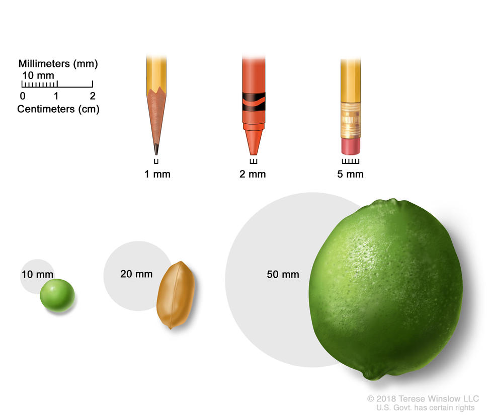
Tumor size is often measured in millimeters (mm) or centimeters. Common items that can be used to show tumor size in mm include: a sharp pencil point (1 mm), a new crayon point (2 mm), a pencil-top eraser (5 mm), a pea (10 mm), a peanut (20 mm), and a lime (50 mm).
Credit: © Terese Winslow
In stage I, cancer has formed and the tumor is 2 centimeters or smaller.
Stage II (also called stage 2) anal cancer
Stage II anal cancer is divided into stages IIA and IIB.
- In stage IIA, the tumor is larger than 2 centimeters but not larger than 5 centimeters.
- In stage IIB, the tumor is larger than 5 centimeters.
Stage III (also called stage 3) anal cancer
Stage III anal cancer is divided into stages IIIA, IIIB, and IIIC.
- In stage IIIA, the tumor is 5 centimeters or smaller and has spread to lymph nodes near the anus or groin.
- In stage IIIB, the tumor is any size and has spread to nearby organs, such as the vagina, urethra, or bladder. Cancer has not spread to lymph nodes.
- In stage IIIC, the tumor is any size and may have spread to nearby organs. Cancer has spread to lymph nodes near the anus or groin.
Stage IV (also called stage 4) anal cancer
In stage IV, the tumor is any size. Cancer may have spread to lymph nodes or nearby organs and has spread to other parts of the body, such as the liver or lungs.
Stage IV anal cancer is also called metastatic anal cancer. Metastatic cancer happens when cancer cells travel through the lymphatic system or blood and form tumors in other parts of the body. The metastatic tumor is the same type of cancer as the primary tumor. For example, if anal cancer spreads to the liver, the cancer cells in the liver are actually anal cancer cells. The disease is called metastatic anal cancer, not liver cancer.
Recurrent anal cancer
Recurrent anal cancer is cancer that has come back after it has been treated. If anal cancer comes back, it may come back in the anus or in other parts of the body, such as the liver or lungs. Tests will be done to help determine where the cancer has returned. The type of treatment for recurrent anal cancer will depend on where it has come back.
Learn more in Recurrent Cancer: When Cancer Comes Back.
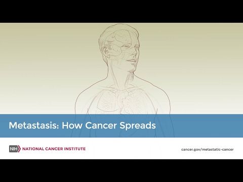
Metastasis: How Cancer Spreads
During metastasis, cancer cells spread from the place in the body where they first formed to other parts of the body.