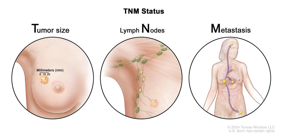
Breast cancer staging uses the TNM (tumor node metastasis) staging system to describe the size of the primary tumor and the spread of cancer to nearby lymph nodes or other parts of the body.
Credit: © Terese Winslow
Tumor (T)
The T category in the TNM staging system describes the size and location of the tumor:
TX: The primary tumor cannot be assessed.
T0: There is no sign of a primary tumor in the breast.
Tis: There is carcinoma in situ in the breast.
- Tis (DCIS): Abnormal cells have been found in the breast ducts and have not spread past the layer of tissue where they began.
- Tis (Paget disease): A rare form of early, noninvasive cancer where abnormal cells are found in the skin of the nipple and have not spread beyond where they first formed. No tumor or DCIS is found in the breast tissue under the nipple.
T1: The tumor is 20 millimeters or smaller. There are four subtypes of a T1 tumor depending on the size of the tumor:
- T1mi: The tumor is 1 millimeter or smaller.
- T1a: The tumor is larger than 1 millimeter but not larger than 5 millimeters.
- T1b: The tumor is larger than 5 millimeters but not larger than 10 millimeters.
- T1c: The tumor is larger than 10 millimeters but not larger than 20 millimeters.
T2: The tumor is larger than 20 millimeters but not larger than 50 millimeters.
T3: The tumor is larger than 50 millimeters.
T4: The tumor is described as one of the following:
- T4a: The tumor has grown into the chest wall.
- T4b: The tumor has grown into the skin. An ulcer has formed on the surface of the skin of the breast, small tumor nodules have formed in the same breast as the primary tumor, and/or the skin of the breast is swollen.
- T4c: The tumor has grown into the chest wall and the skin.
- T4d: One-third or more of the skin on the breast is red and swollen (called peau d’orange). T4d includes inflammatory breast cancer.
Lymph node (N)
This section describes pathological staging of lymph nodes. Pathological staging is done when the lymph nodes are removed by surgery and studied under a microscope by a pathologist. When the lymph nodes are checked using mammography or ultrasound, it is called clinical staging. Clinical staging of lymph nodes is not described here.
The N category describes the location of the lymph nodes where cancer has spread and the size of the cancer in the lymph nodes:
NX: The lymph nodes cannot be assessed.
N0: There is no sign of cancer in the lymph nodes, or there may be tiny clusters of cancer cells not larger than 0.2 millimeters in the lymph nodes.
N1: Cancer is described as one of the following:
- N1mi: Cancer has spread to the axillary (armpit area) lymph nodes and is larger than 0.2 millimeters but not larger than 2 millimeters.
- N1a: Cancer has spread to one to three axillary lymph nodes and the cancer in at least one of the lymph nodes is larger than 2 millimeters.
- N1b: Cancer has spread to lymph nodes near the breastbone on the same side of the body as the primary tumor. The cancer is larger than 0.2 millimeters and is found by sentinel lymph node biopsy. Cancer is not found in the axillary lymph nodes.
- N1c: Cancer has spread to one to three axillary lymph nodes, and the cancer in at least one of the lymph nodes is larger than 2 millimeters. Cancer is also found by sentinel lymph node biopsy in the lymph nodes near the breastbone on the same side of the body as the primary tumor.
N2: Cancer is described as one of the following:
- N2a: Cancer has spread to four to nine axillary lymph nodes, and the cancer in at least one of the lymph nodes is larger than 2 millimeters.
- N2b: Cancer has spread to lymph nodes near the breastbone, and the cancer is found by imaging tests. Cancer is not found in the axillary lymph nodes by sentinel lymph node biopsy or lymph node dissection.
N3: Cancer is described as one of the following:
- N3a: Cancer has spread to 10 or more axillary lymph nodes. The cancer in at least one of the lymph nodes is larger than 2 millimeters, or the cancer has spread to lymph nodes below the collarbone.
- N3b: Cancer is described as one of the following:
- Cancer has spread to one to nine axillary lymph nodes, and the cancer in at least one of the lymph nodes is larger than 2 millimeters. Cancer has also spread to lymph nodes near the breastbone, and the cancer is found by imaging tests.
- Cancer has spread to four to nine axillary lymph nodes, and the cancer in at least one of the lymph nodes is larger than 2 millimeters. Cancer has also spread to lymph nodes near the breastbone on the same side of the body as the primary tumor. The cancer in these lymph nodes is larger than 0.2 millimeters and is found by sentinel lymph node biopsy.
- N3c: Cancer has spread to lymph nodes above the collarbone on the same side of the body as the primary tumor.
Metastasis (M)
The M category describes the spread of cancer to other parts of the body.
M0: There is no sign that cancer has spread to other parts of the body.
M1: Cancer has spread to other parts of the body, most often to the bones, lungs, liver, or brain. If cancer has spread to distant lymph nodes, the cancer in the lymph nodes is larger than 0.2 millimeters. The cancer is called metastatic breast cancer.