Tests to diagnose mesothelioma
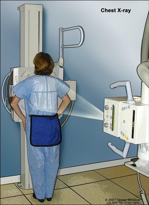
A chest x-ray is used to take pictures of the structures and organs inside the chest. X-rays pass through the patient's body onto film or a computer.
Credit: © Terese Winslow
The tests and procedures used to diagnose mesothelioma may include:
- Chest x-ray is a type of radiation that can go through the body and make pictures of the organs and bones inside the chest.
- CT scan (CAT scan) uses a computer linked to an x-ray machine to make a series of detailed pictures of areas inside the body. The pictures are taken from different angles and are used to create 3-D views of tissues and organs. A dye may be injected into a vein or swallowed to help the organs or tissues show up more clearly. This procedure is also called computed tomography, computerized tomography, or computerized axial tomography. Learn more about Computed Tomography (CT) Scans and Cancer.
- Biopsy is the removal of cells or tissues from the pleura or peritoneum so that a pathologist can view it under a microscope to check for signs of cancer.
Procedures used to collect the cells or tissues include:
- Fine-needle aspiration (FNA) biopsy of the lung uses a thin needle to remove tissue or fluid. An imaging procedure is used to locate the abnormal tissue or fluid in the lung. A small incision may be made in the skin where the biopsy needle is inserted into the abnormal tissue or fluid, and a sample is removed.
- Thoracoscopy is surgery to look at the organs inside the chest to check for abnormal areas. An incision (cut) is made between two ribs, and a thoracoscope (a thin, tube-like instrument with a light and a lens for viewing) is inserted into the chest.
- Thoracotomy is surgery to look inside the chest. An incision (cut) is made between two ribs to check inside the chest for signs of disease.
- Peritoneoscopy is surgery to look inside the abdomen. An incision (cut) is made in the abdominal wall, and a peritoneoscope (a thin, tube-like instrument with a light and a lens for viewing) is inserted into the abdomen to check for signs of disease.
- Open biopsy is surgery to remove abnormal tissues so a pathologist can check it under a microscope for signs of disease. An incision (cut) is made through the skin to expose and remove the tissues.
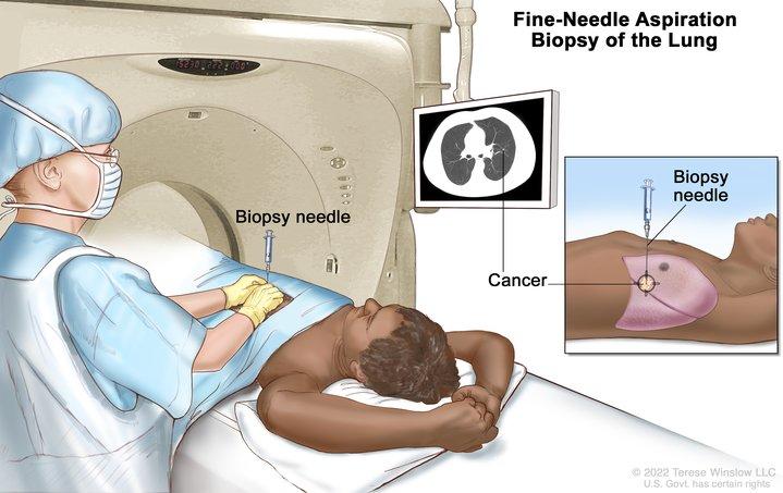
Fine-needle aspiration biopsy of the lung. The patient lies on a table that slides through the computed tomography (CT) machine, which takes x-ray pictures of the inside of the body. The x-ray pictures help the doctor see where the abnormal tissue is in the lung. A biopsy needle is inserted through the chest wall and into the area of abnormal lung tissue. A small piece of tissue is removed through the needle and checked under the microscope for signs of cancer.
Credit: © Terese Winslow
The following tests may be done on the cells and tissue samples that are taken:
- A cytologic exam is a laboratory test to view cells under a microscope to check for anything abnormal. For mesothelioma, fluid is taken from the chest or the abdomen. A pathologist checks the fluid for signs of cancer.
- Immunohistochemistry is a laboratory test that uses antibodies to check for certain antigens (markers) in a sample of a patient's tissue. The antibodies are usually linked to an enzyme or a fluorescent dye. After the antibodies bind to a specific antigen in the tissue sample, the enzyme or dye is activated, and the antigen can then be seen under a microscope. This type of test is used to help diagnose cancer and to help tell one type of cancer from another type of cancer.
- Electron microscopy is a laboratory test in which cells in a sample of tissue are viewed under a high-powered microscope to look for certain changes in the cells. An electron microscope shows tiny details better than other types of microscopes.
Getting a second opinion
You may want to get a second opinion to confirm your cancer diagnosis and treatment plan. If you seek a second opinion, you will need to get medical test results and reports from the first doctor to share with the second doctor. The second doctor will review the pathology report, slides, and scans. They may agree with the first doctor, suggest changes or another treatment approach, or provide more information about your cancer.
Learn more about choosing a doctor and getting a second opinion at Finding Cancer Care. You can contact NCI's Cancer Information Service via chat, email, or phone (both in English and Spanish) for help finding a doctor, hospital, or getting a second opinion. For questions you might want to ask at your appointments, visit Questions to Ask Your Doctor About Cancer.
Mesothelioma prognosis
The prognosis and treatment options depend on:
- the stage of the cancer
- the size of the tumor
- whether the tumor can be removed completely by surgery
- the amount of fluid in the chest or abdomen
- the patient's age
- the patient's activity level
- the patient's general health, including lung and heart health
- the type of mesothelioma cells and how they look under a microscope
- the number of white blood cells and how much hemoglobin is in the blood
- whether the patient is male or female
- whether the cancer has just been diagnosed or has recurred (come back)