Tests to stage mesothelioma
After mesothelioma has been diagnosed, tests are done to find out if cancer cells have spread to other parts of the body.
The process used to find out if cancer has spread outside the pleura or peritoneum is called staging. The information gathered from the staging process determines the stage of the disease. It is important to know whether the cancer has spread in order to plan treatment.
The following tests and procedures may be used in the staging process:
- CT scan (CAT scan) uses a computer linked to an x-ray machine to make a series of detailed pictures of areas inside the body, such as the chest and abdomen. The pictures are taken from different angles and are used to create 3-D views of tissues and organs. A dye may be injected into a vein or swallowed to help the organs or tissues show up more clearly. This procedure is also called computed tomography, computerized tomography, or computerized axial tomography. Learn more at Computed Tomography (CT) Scans and Cancer.
- PET scan (positron emission tomography scan) uses a small amount of radioactive sugar that is injected into a vein. The PET scanner rotates around the body to make detailed, computerized pictures of areas inside the body where the glucose is taken up. Because cancer cells often take up more glucose than normal cells, the pictures can be used to find cancer cells in the body.
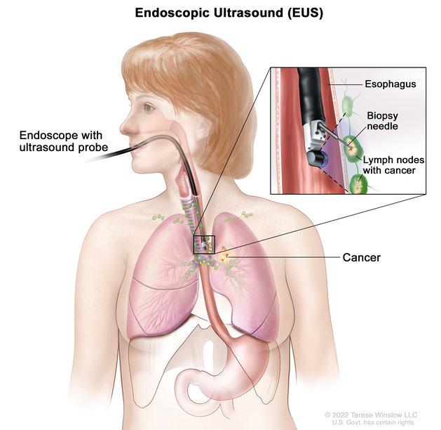
Endoscopic ultrasound-guided fine-needle aspiration biopsy. An endoscope that has an ultrasound probe and a biopsy needle is inserted through the mouth and into the esophagus. The probe bounces sound waves off body tissues to make echoes that form a sonogram (computer picture) of the lymph nodes near the esophagus. The sonogram helps the doctor see where to place the biopsy needle to remove tissue from the lymph nodes. This tissue is checked under a microscope for signs of cancer.
Credit: © Terese Winslow
- MRI (magnetic resonance imaging) uses a magnet, radio waves, and a computer to make a series of detailed pictures of areas inside the body. This procedure is also called nuclear magnetic resonance imaging (NMRI).
- Endoscopic ultrasound (EUS) is a procedure in which an endoscope is inserted into the body. An endoscope is a thin, tube-like instrument with a light and a lens for viewing. A probe at the end of the endoscope is used to bounce high-energy sound waves (ultrasound) off internal tissues or organs and make echoes. The echoes form a picture of body tissues called a sonogram. This procedure is also called endosonography. EUS may be used to guide fine-needle aspiration (FNA) biopsy of the lung, lymph nodes, or other areas.
- Laparoscopy is surgery to look at the organs inside the abdomen to check for signs of disease. Small incisions (cuts) are made in the wall of the abdomen, and a laparoscope (a thin, lighted tube) is inserted into one of the incisions. Other instruments may be inserted through the same or other incisions to perform procedures such as taking tissue samples to be checked under a microscope for signs of disease.
- Lymph node biopsy is the removal of all or part of a lymph node. A pathologist views the lymph node tissue under a microscope to check for cancer cells.
- Mediastinoscopy is surgery to look at the organs, tissues, and lymph nodes between the lungs for abnormal areas. An incision (cut) is made at the top of the breastbone, and a mediastinoscope is inserted into the chest. A mediastinoscope is a thin, tube-like instrument with a light and a lens for viewing. It may also have a tool to remove tissue or lymph node samples, which are checked under a microscope for signs of cancer.
Stage I (also called stage 1) mesothelioma
Stage I is divided into stages IA and IB:
- In stage IA, cancer is found in the inside lining of the chest wall on one side of the chest. On the same side of the chest, cancer may also be found in one or more of the following:
- the thin layer of tissue that covers the lung
- the thin layer of tissue that covers the organs between the lungs
- the thin layer of tissue that covers the top of the diaphragm
- In stage IB, cancer is found in the inside lining of the chest wall, and in each of the thin layers of tissue that cover the lung, the organs between the lungs, and the top of the diaphragm on one side of the chest. On the same side of the chest, cancer has also spread into one or more of the following:
- diaphragm
- lung tissue
- tissue between the ribs and the inside lining of the chest wall
- fat in the area between the lungs
- soft tissues of the chest wall
- sac around the heart
Stage II (also called stage 2) mesothelioma
In stage II, cancer is found in the inside lining of the chest wall on one side of the chest. On the same side of the chest, cancer may also be found in one or more of the following:
- the thin layer of tissue that covers the lung
- the thin layer of tissue that covers the organs between the lungs
- the thin layer of tissue that covers the top of the diaphragm
Cancer has spread to lymph nodes along the center of the chest on the same side of the chest as the tumor.
or
Cancer is found in the inside lining of the chest wall, and in each of the thin layers of tissue that cover the lung, the organs between the lungs, and the top of the diaphragm on one side of the chest. On the same side of the chest, cancer has also spread into one or both of the following:
- diaphragm
- lung tissue
Cancer has spread to lymph nodes along the center of the chest on the same side of the chest as the tumor.
Stage III (also called stage 3) mesothelioma
Stage III is divided into stages IIIA and IIIB.
In stage IIIA, cancer is found in the inside lining of the chest wall, and in each of the thin layers of tissue that cover the lung, the organs between the lungs, and the top of the diaphragm on one side of the chest. On the same side of the chest, cancer has also spread into one or more of the following:
- tissue between the ribs and the inside lining of the chest wall
- fat in the area between the lungs
- soft tissues of the chest wall
- sac around the heart
Cancer has spread to lymph nodes along the center of the chest on the same side of the chest as the tumor.
In stage IIIB, cancer is found in the inside lining of the chest wall, and may also be found in the thin layers of tissue that cover the lung, the organs between the lungs, and/or the top of the diaphragm on one side of the chest. On the same side of the chest, cancer may have also spread into one or more of the following:
- diaphragm
- lung tissue
- tissue between the ribs and the inside lining of the chest wall
- fat in the area between the lungs
- soft tissues of the chest wall
- sac around the heart
Cancer has spread to lymph nodes above the collarbone on either side of the chest or cancer has spread to lymph nodes along the center of the chest on the opposite side of the chest as the tumor.
or
Cancer is found in the inside lining of the chest wall, and in each of the thin layers of tissue that cover the lung, the organs between the lungs, and the top of the diaphragm on one side of the chest. Cancer has also spread to one or more of the following:
- the chest wall and may be found in the rib
- through the diaphragm into the peritoneum
- the tissue lining the chest on the opposite side of the body as the tumor
- the organs in the area between the lungs (esophagus, trachea, thymus, blood vessels)
- the spine
- through the sac around the heart or into the heart muscle
Cancer may have spread to lymph nodes.
Stage IV (also called stage 4) mesothelioma
In stage IV, cancer has spread to the tissue covering the lung or the lung on the opposite side of the chest, peritoneum, bones, liver, lymph nodes outside the chest, or to other parts of the body.
Stage IV mesothelioma is also called metastatic mesothelioma. Metastatic cancer happens when cancer cells travel through the lymphatic system or blood and form tumors in other parts of the body. The metastatic tumor is the same type of cancer as the primary tumor. For example, if mesothelioma spreads to the liver, the cancer cells in the liver are actually mesothelioma cells. The disease is called metastatic mesothelioma, not liver cancer.
Recurrent mesothelioma
Recurrent mesothelioma is cancer that has come back after it has been treated. If mesothelioma comes back, it may come back in the chest or in other parts of the body, such as the liver, lungs, or both. Tests will be done to help determine where the cancer has returned. The type of treatment for recurrent mesothelioma will depend on where it has come back.
Learn more in Recurrent Cancer: When Cancer Comes Back.