Making sense of test results
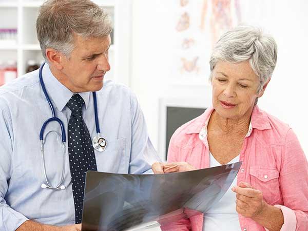
Results from lab tests, imaging, and biopsies are often posted in your patient portal before your doctor can discuss them with you. It is normal to feel anxious and want to know right away what the results are and what they mean. But your doctor is the best person to explain the results from all your tests and what they mean for you.
Lab tests used to diagnose cancer
High or low levels of certain substances in your body can be a sign of cancer. So lab tests of your blood, urine, and other body fluids that measure these substances can help doctors make a diagnosis. However, abnormal lab results are not a sure sign of cancer. Lab test results are used along with the results of other tests, such as biopsies and imaging, to help diagnose and learn more about a person’s cancer.
It is important to keep in mind that lab results for healthy people can vary from person to person. Reasons for these differences include age, sex, race, medical history, and general health. In fact, your own results can vary from day to day. Because normal results can bounce around a bit, they are often reported in a range, with lower and upper limits. These ranges are based on test results from large numbers of people who have been tested in the past.
For many tests, it is possible to have normal results even if you have cancer. And it is possible to have test results outside the normal range even if you are healthy. These are some of the reasons why lab tests alone can’t say for sure if you have cancer or any other disease.
Your doctor is the best person to explain your lab test results and what they mean for you.
Some common types of lab tests used to help diagnose cancer are listed below.
Blood chemistry test
What it does: A blood chemistry test measures the amounts of certain substances that are released into the blood by the organs and tissues of the body. These substances include metabolites, electrolytes, fats, sugars, and proteins, including enzymes.
What it tells us: Blood chemistry tests give important information about how well your kidneys, liver, and other organs are working. High or low levels of some substances in the blood can be a sign of disease or treatment side effects.
Complete blood count (CBC)
What it does: The CBC measures the number of red blood cells, white blood cells, and platelets in your blood. It also measures the amount of hemoglobin (the protein that carries oxygen) in your blood, the amount of your blood that is made up of red blood cells (hematocrit), the size of your red blood cells, and the amount of hemoglobin in your red blood cells.
How it is used: The CBC is often part of a routine health check-up. It can help diagnose some cancers, especially leukemias. It is also used to monitor your health during and after treatment.
Cytogenetic analysis
What it does: Cytogenetic analysis looks for changes in chromosomes in samples of tissue, blood, bone marrow, or amniotic fluid. Chromosome changes may include broken, missing, rearranged, or extra chromosomes. Changes in certain chromosomes may be a sign of a genetic condition or some types of cancer.
How it is used: Cytogenetic analysis may be used to help diagnose cancer, plan treatment, or find out how well treatment is working.
Immunophenotyping
What it does: Immunophenotyping uses antibodies to identify cells based on the types of antigens or markers on the surface of the cells. It is most often done on blood or bone marrow samples. But it may also be done on other body fluids or tissue samples.
How it is used: Immunophenotyping helps diagnose, stage, and monitor blood cancers and other blood disorders, such as leukemias, lymphomas, myelodysplastic syndromes, and myeloproliferative disorders.
Liquid biopsy
What it does: Liquid biopsy is a test done on a sample of blood to look for cancer cells or pieces of DNA from tumor cells that are sometimes released into the blood.
How it is used: A liquid biopsy may help find cancer at an early stage. It may also be used to help plan treatment or to find out how well treatment is working or if cancer has come back.
Sputum cytology
What it does: Sputum cytology looks for abnormal cells in sputum, which is mucus and other matter brought up from the lungs by coughing.
How it is used: Sputum cytology can help diagnose lung cancer.
Tumor marker tests
What they do: Tests for tumor markers measure substances that are produced by cancer cells or other cells of the body in response to cancer. Most tumor markers are made by both normal cells and cancer cells. But they are produced at much higher levels by cancer cells.
How they are used: Tumor markers can be used to help diagnose cancer, decide on treatment, measure how well treatment worked, and watch for signs that the cancer has returned.
Learn more about tumor markers.
Urinalysis
What it does: Urinalysis describes the color of urine and measures its contents, such as sugar, protein, red blood cells, and white blood cells.
How it is used: Urinalysis can help diagnose kidney cancer, bladder cancer, and rarer urothelial cancers.
Urine cytology
What it does: Urine cytology finds disease by looking for abnormal cells shed from the urinary tract into the urine.
How it is used: Urine cytology helps diagnose kidney cancer, bladder cancer, and rarer urothelial cancers. After cancer treatment, it is used to watch for signs that cancer has returned.
Imaging tests used in cancer
Imaging tests create pictures of areas inside your body that help the doctor see whether a tumor is present. These pictures can be made in several ways.
CT scan
A CT scan uses an x-ray machine linked to a computer to take a series of pictures of your organs from different angles. These pictures are used to create detailed 3-D images of the inside of your body.
Sometimes, you may receive a dye or other contrast material before you have the scan. You might swallow the dye, or it may be given by a needle into a vein. Contrast material helps make the pictures easier to read by highlighting certain areas in the body.
During the CT scan, you will lie still on a table that slides into a donut-shaped scanner. The CT machine moves around you, taking pictures.
Learn more about CT scans and how they are used to diagnose cancer.
MRI
An MRI uses a powerful magnet and radio waves to take pictures of your body in slices. These slices are combined to create detailed images of the inside of your body, which can show places where there may be tumors.
When you have an MRI, you lie still on a table that is pushed into a long chamber that surrounds part or all of your body. The MRI machine makes loud thumping noises and rhythmic beats.
Sometimes, you might have a special dye injected into your vein before or during your MRI exam. This dye, called a contrast agent, can make tumors show up brighter in the pictures.
Nuclear scan
A nuclear scan uses radioactive material to take pictures of the inside of the body. This type of scan may also be called a radionuclide scan.
Before this scan, you receive an injection of a small amount of radioactive material, which is sometimes called a tracer. It flows through your bloodstream and collects in certain bones or organs.
During the scan, you lie still on a table while a machine called a scanner detects and measures the radioactivity in your body, creating pictures of bones or organs on a computer screen or on film.
After the scan, the radioactive material in your body will lose its radioactivity over time. It may also leave your body through your urine or stool.
Bone scan
Bone scans are a type of nuclear scan that check for abnormal areas or damage in the bones. They may be used to diagnose bone cancer or find out whether cancer has spread to the bones from elsewhere in the body (called metastatic bone tumors).
Before this test, a very small amount of radioactive material is injected into your vein. As it travels through the blood, the material collects in abnormal areas in the bone. Areas where the material collects show up on pictures taken by a special scanner. These areas are called “hot spots.”
PET scan
A PET scan is a type of nuclear scan that makes detailed 3-D pictures of areas inside your body where glucose is taken up. Because cancer cells often take up more glucose than healthy cells, the pictures can be used to find cancer in the body.
Before the scan, you receive an injection of a tracer called radioactive glucose. During the scan, you will lie still on a table that moves back and forth through a scanner.
Ultrasound
An ultrasound exam uses high-energy sound waves that people cannot hear. The sound waves echo off tissues inside your body. A computer uses these echoes to create pictures of areas inside your body. This picture is called a sonogram.
During an ultrasound exam, you will lie on a table while a technologist slowly moves a device called a transducer, which makes the high-energy sound waves, on the skin over the part of the body that is being examined. The transducer is covered with a warm gel that helps it glide over the skin.
X-rays
X-rays use low doses of radiation to create pictures inside your body. An x-ray technologist will put you in position and direct the x-ray beam to the correct part of your body. While the images are taken, you will need to stay very still and may need to hold your breath for a second or two.
Biopsy
In most cases, doctors need to do a biopsy to be certain that you have cancer. A biopsy is a procedure in which the doctor removes a sample of abnormal tissue. A pathologist looks at the tissue under a microscope and runs other tests on the cells in the sample. The pathologist describes the findings in a pathology report, which contains details about your diagnosis. The information in your pathology reports can also help show what treatment options might work for you.
Learn more about pathology reports and the type of information they contain.
The biopsy sample may be obtained in several ways.
With a needle: The doctor uses a needle to withdraw tissue or fluid. This method is used for bone marrow aspirations, spinal taps, and some breast, prostate, and liver biopsies.
With endoscopy: The doctor inserts a thin, lighted tube called an endoscope into a natural body opening, such as the mouth or anus. The doctor can remove some or all of the abnormal tissue through the endoscope.
Examples of endoscopy exams include:
- Colonoscopy, which is an exam of the colon and rectum. In this type of exam, an endoscope goes through the anus.
- Bronchoscopy, which is an exam of the trachea, bronchi, and lungs. In this type of exam, an endoscope goes through the mouth or nose and down the throat.
With surgery: A surgeon removes an area of abnormal cells during an operation. Surgery may be excisional or incisional.
In an excisional biopsy, the surgeon removes the entire area of abnormal cells. Often some of the normal tissue around these cells is also removed.
In an incisional biopsy, the surgeon removes just part of the abnormal area.
Some biopsies may require a sedative or anesthesia.
Sedatives are medicine that helps you relax and stay very still or sleep during a biopsy.
Anesthesia keeps you from feeling pain. It refers to drugs or other substances that cause you to lose feeling or awareness. There are three types of anesthesia.
- local anesthesia, which causes loss of feeling in one small area of the body
- regional anesthesia, which causes loss of feeling in a part of the body, such as an arm or leg
- general anesthesia, which causes loss of feeling and a complete loss of awareness that seems like a very deep sleep
What happens if tests show you have cancer
If the biopsy and other tests show that you have cancer, you may have more tests to help your doctor plan treatment. For instance, your doctor may do other tests to figure out if the cancer has spread and how far. This information is important for knowing the stage of your cancer. For some cancers, other pathology studies are done to find out the grade of the tumor. Or tumor markers are studied to find out the risk group that you fall into. This information is important for deciding on the best treatment. Your tumor may also be tested further for other tumor markers or biomarkers.
To learn more about the diagnosis, staging, and treatment for your type of cancer, see the PDQ cancer treatment summaries for adult and childhood cancers.