Risk factors for childhood adrenocortical carcinoma
Anything that increases your chance of getting a disease is called a risk factor. Having a risk factor does not mean that you will get cancer; not having risk factors doesn't mean that you will not get cancer. Talk with your child's doctor if you think your child may be at risk.
The risk of adrenocortical carcinoma is increased by having a mutation (change) in the TP53 gene or any of the following syndromes:
Symptoms of childhood adrenocortical carcinoma
These and other signs and symptoms may be caused by adrenocortical carcinoma or by other conditions.
Check with your child's doctor if your child has any of the following:
- Pain in the abdomen or back.
- A lump in the abdomen.
- Feeling of fullness in the abdomen.
- High blood pressure.
- Acne.
- Growing body hair.
- Deepening of the voice.
- Growing faster than normal.
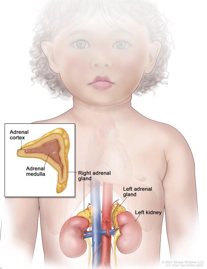
Also, cancer of the adrenal cortex may be functioning (makes more hormones than normal) or nonfunctioning (does not make extra hormones). Most tumors of the adrenal cortex in children are functioning tumors. The extra hormones made by functioning tumors may cause certain signs or symptoms of disease and these depend on the type of hormone made by the tumor. For example, extra androgen hormone may also cause male children to develop an enlarged penis and female children to develop enlarged genitalia. Extra estrogen hormone may cause the growth of breast tissue in male children. Extra cortisol hormone may cause (hypercortisolism).
Learn more about Adrenocortical Carcinoma Symptoms.
Tests to diagnose childhood adrenocortical carcinoma
Tests are done to diagnose and stage cancer. After cancer is diagnosed, more tests are done to find out if cancer cells have spread to nearby areas or to other parts of the body. This process is called staging. It is important to know whether cancer has spread in order to plan the best treatment.
The following tests and procedures may be used:
- Physical exam and health history: An exam of the body to check general signs of health, including checking for signs of disease, such as lumps or anything else that seems unusual. A history of the patient’s health habits and past illnesses and treatments will also be taken.
- X-ray: An x-ray of the chest, abdomen, or bones inside the body. An x-ray is a type of energy beam that can go through the body and onto film, making a picture of areas inside the body.
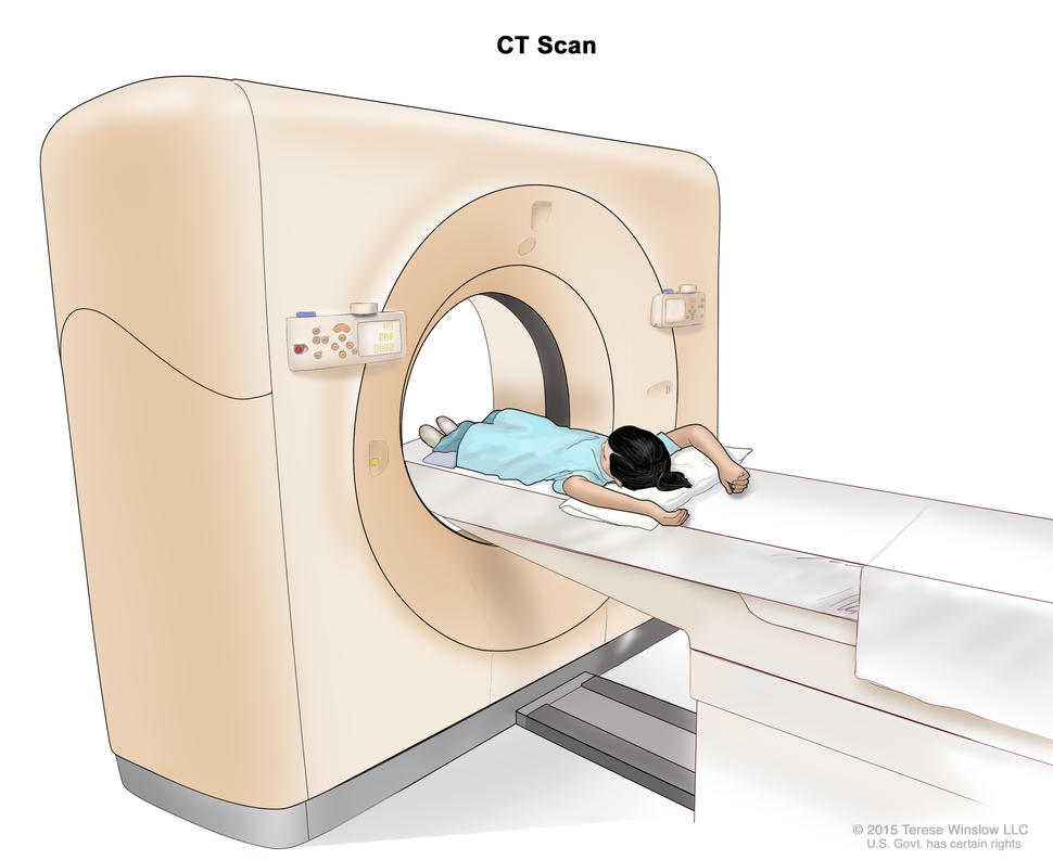
Computed tomography (CT) scan. The child lies on a table that slides through the CT scanner, which takes a series of detailed x-ray pictures of areas inside the body.
Credit: © Terese Winslow
- CT scan: A procedure that makes a series of detailed pictures of areas inside the body, such as the chest or abdomen, taken from different angles. The pictures are made by a computer linked to an x-ray machine. A dye may be injected into a vein or swallowed to help the organs or tissues show up more clearly. This procedure is also called computed tomography, computerized tomography, or computerized axial tomography.
- MRI (magnetic resonance imaging): A procedure that uses a magnet, radio waves, and a computer to make a series of detailed pictures of areas of the body, such as the chest and abdomen. This procedure is also called nuclear magnetic resonance imaging (NMRI).
- PET scan: A procedure to find malignant tumor cells in the body. A small amount of radioactive glucose (sugar) is injected into a vein. The PET scanner rotates around the body and makes a picture of where glucose is being used in the body. Malignant tumor cells show up brighter in the picture because they are more active and take up more glucose than normal cells do.
- Ultrasound exam: A procedure in which high-energy sound waves (ultrasound) are bounced off internal tissues or organs, such as the abdomen, and make echoes. The echoes form a picture of body tissues called a sonogram. The picture can be printed to be looked at later.
- Biopsy: The removal of cells or tissues during surgery so they can be viewed under a microscope by a pathologist to check for signs of cancer.
- Blood chemistry studies: A procedure in which a blood sample is checked to measure the amounts of certain substances released into the blood by organs and tissues in the body. An unusual (higher or lower than normal) amount of a substance can be a sign of disease.
- Blood hormone studies: A procedure in which a blood sample is checked to measure the amounts of certain hormones released into the blood by organs and tissues in the body. An unusual (higher or lower than normal) amount of a substance can be a sign of disease in the organ or tissue that makes it. The blood may be checked for testosterone or estrogen. A higher-than-normal amount of these hormones may be a sign of adrenocortical carcinoma.
- Twenty-four-hour urine test: A test in which urine is collected for 24 hours to measure the amounts of cortisol or 17-ketosteroids. A higher-than-normal amount of these substances in the urine may be a sign of disease in the adrenal cortex.
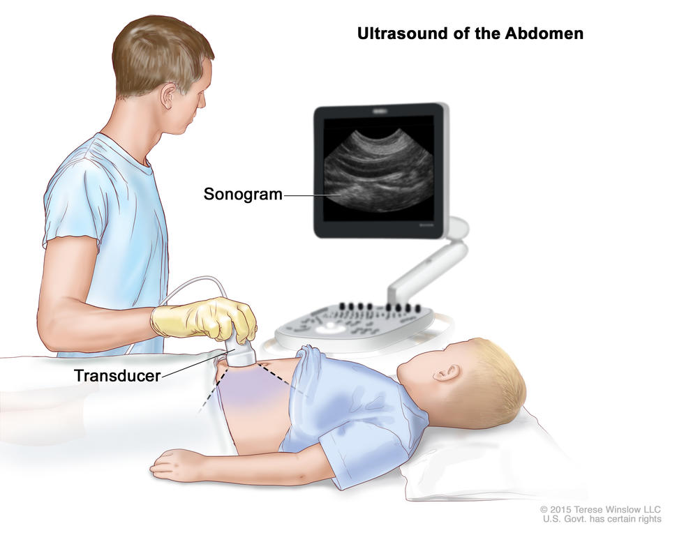
Abdominal ultrasound. An ultrasound transducer connected to a computer is pressed against the skin of the abdomen. The transducer bounces sound waves off internal organs and tissues to make echoes that form a sonogram (computer picture).
Credit: © Terese Winslow
- Low-dose dexamethasone suppression test: A test in which one or more small doses of dexamethasone are given. The level of cortisol is checked from a sample of blood or from urine that is collected for three days. This test is done to check if the adrenal gland is making too much cortisol.
- High-dose dexamethasone suppression test: A test in which one or more high doses of dexamethasone are given. The level of cortisol is checked from a sample of blood or from urine that is collected for three days. This test is done to check if the adrenal gland is making too much cortisol or if the pituitary gland is telling the adrenal glands to make too much cortisol.
- Adrenal angiography: A procedure to look at the arteries and the flow of blood near the adrenal gland. A contrast dye is injected into the adrenal arteries. As the dye moves through the blood vessel, a series of x-rays are taken to see if any arteries are blocked.
- Adrenal venography: A procedure to look at the adrenal veins and the flow of blood near the adrenal glands. A contrast dye is injected into an adrenal vein. As the contrast dye moves through the vein, a series of x-rays are taken to see if any veins are blocked. A catheter (very thin tube) may be inserted into the vein to take a blood sample, which is checked for abnormal hormone levels.
Prognostic factors for childhood adrenocortical carcinoma
The prognosis is good for patients who have small tumors that have been completely removed by surgery. For other patients, the prognosis depends on the following:
- The size of the tumor.
- How quickly the cancer is growing.
- Whether there are changes in certain genes.
- Whether the tumor has spread to other parts of the body, including the lymph nodes, liver, lung, kidney, or bone.
- The child's age.
- Whether the covering around the tumor broke open during surgery to remove the tumor.
- Whether the tumor was completely removed during surgery.
- Whether the child has developed masculine traits.
Stages of childhood adrenocortical carcinoma
The process used to find out if cancer has spread to tissues near the adrenal glands or to other parts of the body is called staging. The information gathered from the staging process is used to plan treatment. The results of the tests and procedures used to diagnose cancer are often also used to stage the disease.
Sometimes childhood adrenocortical carcinoma recurs (comes back) in the adrenal cortex or in other parts of the body after it has been treated.
There are three ways that cancer spreads in the body
Cancer can spread through tissue, the lymph system, and the blood:
- Tissue. The cancer spreads from where it began by growing into nearby areas.
- Lymph system. The cancer spreads from where it began by getting into the lymph system. The cancer travels through the lymph vessels to other parts of the body.
- Blood. The cancer spreads from where it began by getting into the blood. The cancer travels through the blood vessels to other parts of the body.
Cancer may spread from where it began to other parts of the body
When cancer spreads to another part of the body, it is called metastasis. Cancer cells break away from where they began (the primary tumor) and travel through the lymph system or blood.
- Lymph system. The cancer gets into the lymph system, travels through the lymph vessels, and forms a tumor (metastatic tumor) in another part of the body.
- Blood. The cancer gets into the blood, travels through the blood vessels, and forms a tumor (metastatic tumor) in another part of the body.
The metastatic tumor is the same type of cancer as the primary tumor. For example, if adrenocortical carcinoma spreads to the liver, the cancer cells in the liver are actually adrenocortical carcinoma cells. The disease is metastatic adrenocortical carcinoma, not liver cancer.
Metastasis: How Cancer Spreads
During metastasis, cancer cells spread from the place in the body where they first formed to other parts of the body.
Treatment options for children with adrenocortical carcinoma
There are different types of treatment for children with adrenocortical carcinoma.
Some treatments are standard (the currently used treatment), and some are being tested in clinical trials. A treatment clinical trial is a research study meant to help improve current treatments or obtain information on new treatments for patients with cancer. When clinical trials show that a new treatment is better than the standard treatment, the new treatment may become the standard treatment.
Because cancer in children is rare, taking part in a clinical trial should be considered. Some clinical trials are open only to patients who have not started treatment.
Who treats children with adrenocortical carcinoma?
Treatment will be overseen by a pediatric oncologist, a doctor who specializes in treating children with cancer. The pediatric oncologist works with other pediatric health professionals who are experts in treating children with cancer and who specialize in certain areas of medicine. This may include the following specialists and others:
- Pediatrician.
- Pediatric surgeon.
- Radiation oncologist.
- Pathologist.
- Endocrinologist.
- Pediatric nurse specialist.
- Social worker.
- Rehabilitation specialist.
- Psychologist.
- Child-life specialist.
Two types of standard treatment are used:
Surgery
Surgery to remove the tumor is the main treatment for adrenocortical carcinoma.
Chemotherapy
Chemotherapy is a cancer treatment that uses drugs to stop the growth of cancer cells, either by killing the cells or by stopping them from dividing. When chemotherapy is taken by mouth or injected into a vein or muscle, the drugs enter the bloodstream and can reach cancer cells throughout the body (systemic chemotherapy).
New treatments being tested in clinical trials
This summary section describes treatments that are being studied in clinical trials. It may not mention every new treatment being studied. Information about clinical trials is available from the NCI website.
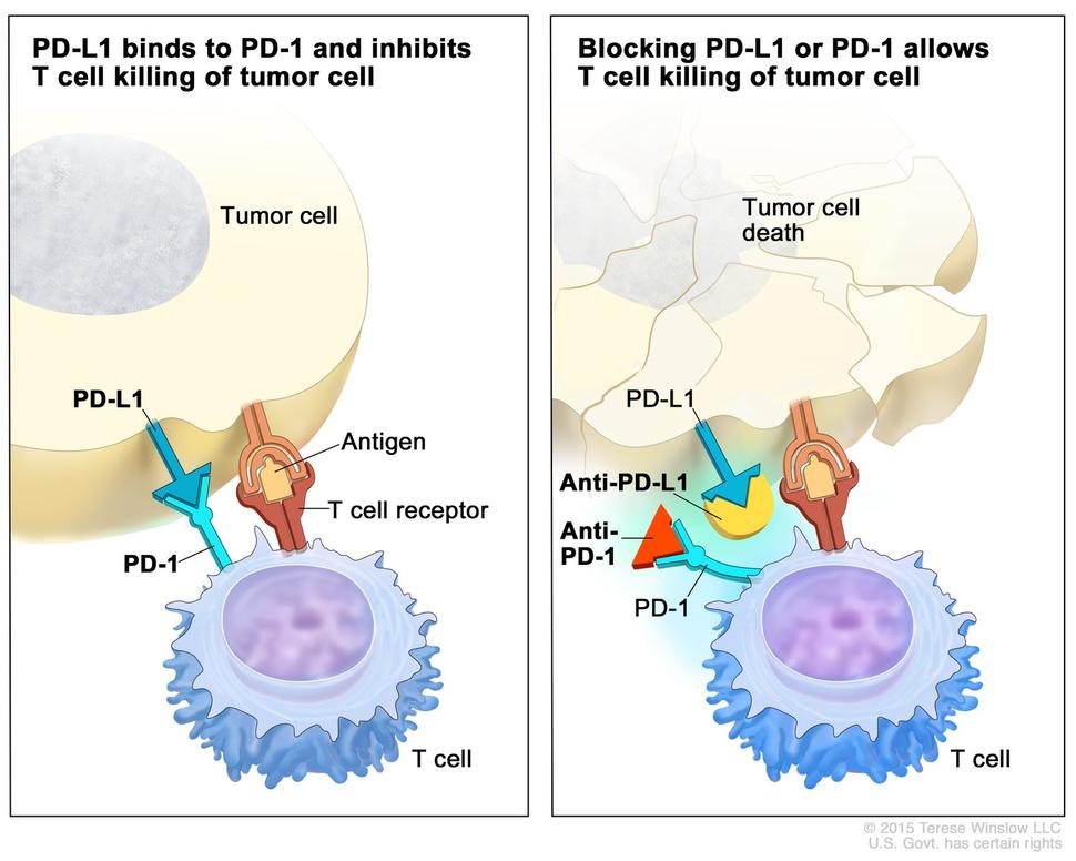
Checkpoint proteins, such as PD-L1 on tumor cells and PD-1 on T cells, help keep immune responses in check. The binding of PD-L1 to PD-1 keeps T cells from killing tumor cells in the body (left panel). Blocking the binding of PD-L1 to PD-1 with an immune checkpoint inhibitor (anti-PD-L1 or anti-PD-1) allows the T cells to kill tumor cells (right panel).
Credit: © Terese Winslow
Immunotherapy
Immunotherapy is a treatment that uses the patient’s immune system to fight cancer. Substances made by the body or made in a laboratory are used to boost, direct, or restore the body’s natural defenses against cancer.
- Immune checkpoint inhibitor therapy is a type of immunotherapy that blocks certain proteins. PD-1 is a protein on the surface of T cells that helps keep the body’s immune responses in check. PD-L1 is a protein found on some types of cancer cells. When PD-1 attaches to PD-L1, it stops the T cell from killing the cancer cell. PD-1 and PD-L1 inhibitors keep PD-1 and PD-L1 proteins from attaching to each other. This allows the T cells to kill cancer cells. Pembrolizumab is a PD-1 inhibitor that is being studied in the treatment of childhood adrenocortical carcinoma that is advanced or has come back after treatment.
Immune Checkpoint Inhibitors
Learn about immune checkpoint inhibitors, one type of immunotherapy used to treat cancer.
Treatment of newly diagnosed childhood adrenocortical carcinoma
Treatment of newly diagnosed adrenocortical carcinoma in children may include the following:
- Surgery to remove the adrenal gland and, if needed, cancer that has spread to other parts of the body. Sometimes chemotherapy is also given.
Treatment of recurrent childhood adrenocortical carcinoma
Treatment of recurrent adrenocortical carcinoma in children may include the following:
Clinical trials
For some patients, taking part in a clinical trial may be the best treatment choice. Clinical trials are part of the cancer research process. Clinical trials are done to find out if new cancer treatments are safe and effective or better than the standard treatment.
Many of today's standard treatments for cancer are based on earlier clinical trials. Patients who take part in a clinical trial may receive the standard treatment or be among the first to receive a new treatment.
Patients who take part in clinical trials also help improve the way cancer will be treated in the future. Even when clinical trials do not lead to effective new treatments, they often answer important questions and help move research forward.
Patients can enter clinical trials before, during, or after starting their cancer treatment
Some clinical trials only include patients who have not yet received treatment. Other trials test treatments for patients whose cancer has not gotten better. There are also clinical trials that test new ways to stop cancer from recurring (coming back) or reduce the side effects of cancer treatment.
Clinical trials are taking place in many parts of the country. Find clinical trials for adrenocortical carcinoma at Treatment Clinical Trials for Adrenal Cortex Cancer. Clinical trials supported by other organizations can be found on the ClinicalTrials.gov website. General information about clinical trials is also available.
Side effects of treatment
To learn more about side effects that begin during treatment for cancer, visit Side Effects.
Side effects from cancer treatment that begin after treatment and continue for months or years are called late effects. Late effects of cancer treatment may include the following:
- Physical problems, including problems with the thyroid gland, hearing, fully opening the mouth, dental cavities, and chronic sinusitis or ear infections.
- Changes in mood, feelings, thinking, learning, or memory.
- Second cancers (new types of cancer) or other conditions.
Some late effects may be treated or controlled. It is important to talk with your child's doctors about the possible late effects caused by some treatments. See the PDQ summary on Late Effects of Treatment for Childhood Cancer for more information.
Follow-up care
As your child goes through treatment, they will have follow-up tests or check-ups. Some tests that were done to diagnose or stage the cancer may be repeated to see how well the treatment is working. Decisions about whether to continue, change, or stop treatment may be based on the results of these tests.
Some of the tests will continue to be done from time to time after treatment has ended. The results of these tests can show if your child's condition has changed or if the cancer has recurred (come back).