
Biology of Cancer - Cancer Currents Blog
Cancer biology research news, with context from experts at NCI and elsewhere. Topics include cancer metastasis, the tumor microenvironment, and new targets for cancer therapies.
-
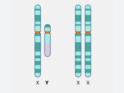
In men, loss of the Y chromosome in bladder cancer cells helped tumors evade the immune system and grow unchecked, a new study shows. However, losing the chromosome also appears to make bladder cancer more susceptible to immunotherapy, researchers reported.
-
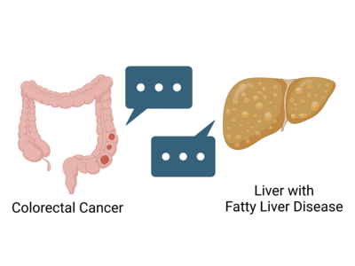
When colorectal cancer spreads to the liver, it can be very difficult to treat. Cancer is more likely to invade the liver when patients have fatty liver disease. A recent study places the blame on “message bubbles” called extracellular vesicles that are released by the liver.
-
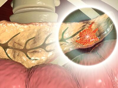
A new study finds that pancreatic cancer cells have a ready way to overcome a lack of glucose, a frequent occurrence in this disease. They use another fuel source: a molecule called uridine. Findings from a related study suggest other cancers do as well.
-
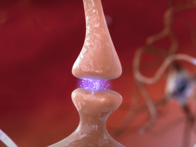
Researchers have found that the aggressive brain cancer glioblastoma can co-opt the formation of new synapses to fuel its own growth. This neural redirection also appears to play a role in the devastating cognitive decline seen in many people with glioblastoma.
-

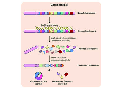
A new study shows for the first time that a circular form of DNA, called ecDNA, is present in precancerous tissue and not just cancer cells. The study also suggested that when ecDNA is present in Barrett’s esophagus, that tissue is almost certain to become cancer.
-
Researchers have identified a mechanism by which cancer cells develop specific genetic changes needed to become resistant to targeted therapies. They also showed that this process, called non-homologous end-joining (NHEJ), can potentially be disrupted.
-
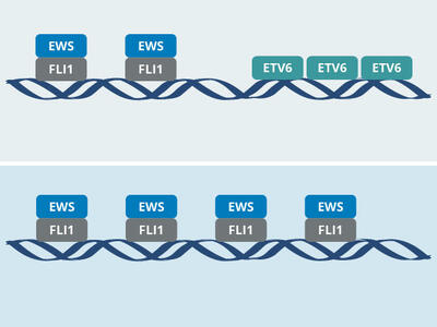
The protein ETV6 appears to promote tumor growth by affecting the behavior of the EWS-FLI1 fusion protein that drives most Ewing sarcomas. The research groups that made the discovery hope it leads to a targeted therapy for the aggressive childhood cancer.
-
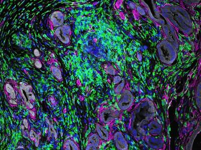
An experimental drug, MRTX1133, shrank tumors or halted their growth in several mouse models of human pancreatic cancer with KRAS G12D mutations, a new study shows. The models included one that is genetically engineered to closely mimic the human disease.
-
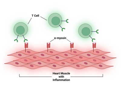
In people with cancer treated with immune checkpoint inhibitors, a rare, but often fatal, side effect is inflammation in the heart, called myocarditis. Researchers have now identified a potential chief cause of this problem: T cells attacking a protein in heart cells called α-myosin.
-
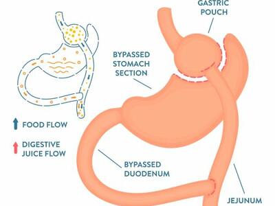
A new study suggests that people with obesity who had bariatric surgery had a much lower risk of five common cancers that aren’t related to hormone levels, including lung, colorectal, and esophageal cancer.