Gliomatosis cerebri is a primary central nervous system (CNS) tumor. This means it begins in the brain or spinal cord.
This tumor is no longer recognized as a formal diagnosis. Instead, gliomatosis cerebri refers to a diffuse pattern of glioma cells with extensive growth that invade multiple lobes of the brain. Gliomas of different grades and cells of origin (such as astrocytes or oligodendrocytes) can grow with this pattern. Very little is currently understood about the molecular basis of the disease. More research is needed to discover the origin of these tumors and improve treatment.
To get an accurate diagnosis, a piece of tumor tissue will be removed during surgery, if possible. A neuropathologist should then review the tumor tissue.
What Are the Grades of Gliomatosis Cerebri?
Primary CNS tumors are graded based on a tumor tissue analysis performed by a neuropathologist.
Gliomatosis cerebri is no longer recognized as a separate entity. Rather, it is thought to be comprised of different types of primary brain tumors with varying grades. As such, the grading of gliomatosis cerebri is based on the grade of the underlying tumor examined after a biopsy.
- Grade 2 (also written as grade II) tumors are low-grade, slow-growing tumors that are more amenable to treatment.
- Grades 3 and 4 (also written as grades III and IV) tumors are high-grade, fast-growing tumors that are more resistant to treatment.
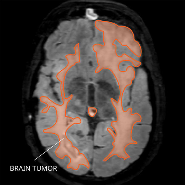
What Does Gliomatosis Cerebri Look like on an MRI?
Gliomatosis cerebri usually appears abnormal in three or more lobes of the brain on a magnetic resonance imaging (MRI) scan.
- Type 1 shows a widespread tumor pattern or a fluffy looking abnormality but no obvious mass.
- Type 2 shows a widespread fluffy looking tumor pattern, as well as a tumor mass.
Finding a gliomatosis cerebri on an MRI does not reliably predict the underlying tumor diagnosis based on tissue findings.

What Causes Gliomatosis Cerebri?
Cancer is a genetic disease—that is, it is caused by certain changes to genes that control the way our cells function. Genes may be mutated (changed) in many types of cancer, which can increase the growth and spread of cancer cells. The cause of gliomatosis cerebri is not known.
Where Does Gliomatosis Cerebri Form?
Gliomatosis cerebri commonly arises from glial cells in the brain. Glial cells support and insulate neurons and are the most abundant cells in the brain. Under a microscope, the tumor cells resemble glial cells and involve many areas of the cerebrum. That is where gliomatosis cerebri gets its name.
Does Gliomatosis Cerebri Spread?
Gliomatosis cerebri can spread to other areas of the CNS through direct extension. They can spread quickly and deeply into surrounding brain tissue. Only rarely do they spread into the spine or by cerebrospinal fluid (CSF). They do not spread outside the CNS.
What Are the Symptoms of Gliomatosis Cerebri?
Gliomatosis cerebri symptoms depend on the tumor’s location and person’s age. Here are some possible symptoms that can occur:
- Seizures
- Fatigue
- Mood changes
- Changes in thinking and memory
- Headaches
Who Is Diagnosed with Gliomatosis Cerebri?
Gliomatosis cerebri is most common in older adults but can occur at any age. Gliomatosis cerebri occurs slightly more often in males than females. An estimated 80 people are living with this tumor in the United States.
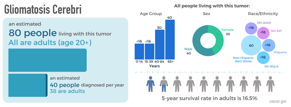
The blue box above shows the estimated number of people living with this disease (prevalence) as of December 31, 2019, as well as the average newly diagnosed cases per year (2008-2019) across all ages and adults (20+ years) specifically. The donut chart shows prevalence by sex; the bar graph shows prevalence by age group; and the packed circles chart shows prevalence by race and Hispanic ethnicity (American Indian/Alaska Native [AIAN]; Asian/Pacific Islander [API]). The people icons show the five-year relative survival rate in adults (2008-2018).
What Is the Prognosis of Gliomatosis Cerebri?
The likely outcome of the disease or chance of recovery is called prognosis. Prognosis is based on tumor grade, location, tumor type, extent of tumor spread, genetic findings, the patient’s age, and tumor remaining after surgery (if surgery is possible).
The relative five-year survival rate for gliomatosis cerebri is 16.5 percent. However, many factors can affect prognosis. These include the tumor grade and molecular type, the person’s age and health when diagnosed, and how they respond to treatment. If you want to understand your prognosis, talk to your doctor.
What Are the Treatment Options for Gliomatosis Cerebri?
The first treatment for a gliomatosis cerebri is surgery, if possible. The goal of surgery is to obtain tissue to determine the tumor type and remove as much of the tumor as possible without causing more symptoms. Surgery is usually limited to a biopsy, as there is not a central mass for removal.
After surgery, there is no standard treatment for gliomatosis cerebri. Other treatments may include radiation, chemotherapy, or clinical trials. Gliomatosis cerebri may respond well to radiation therapy, but patients should discuss the full risks and benefits with their health care team, as radiation to a large volume of the brain puts normal brain tissue at risk. Chemotherapy is also a treatment that may be recommended during radiation, after radiation is completed, or if the tumor recurs after initial treatment. Temozolomide is a drug that is often used, as this is the therapy for astrocytic type tumors. Clinical trials testing new chemotherapy, targeted therapy, or immunotherapy drugs may also be available.
Open Clinical Studies for Gliomatosis Cerebri
- PLX038 in CNS Tumors
- Immune Checkpoint Inhibitor Nivolumab for Patients with Rare CNS Cancers
- Nivolumab for Patients with IDH-Mutant Gliomas
- ONC206 for Patients with Rare CNS Neoplasms
Learn More
- Video: Clinical Trial Tests Nivolumab for Patients with Rare Brain and Spine Cancers
- Statistical Report Highlights Key Trends in Adolescents and Young Adults with Brain Tumors
- Modifying a Chemotherapy Drug Offers Hope to People with Rare Brain and Spine Tumors
- Smart Wearables Show Promise for Tracking Sleep Patterns in Brain Tumor Patients
- Read our NCI-CONNECTions Blog for current news and information on brain and spine tumors

Referrals
NCI-CONNECT doctors and nurses work with you and your primary doctor to collaborate on a comprehensive care plan that treats your brain or spine tumor. They will also help you cope with the physical and emotional aspects of your diagnosis. Learn about requesting a consultation >